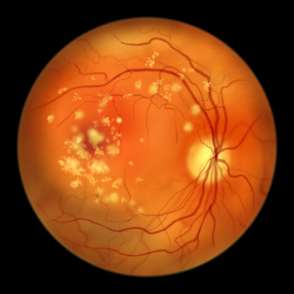
Drusen (singular: druse) are small yellow deposits of protein and lipids (fat) that develop under the retina. The retina is the light-sensitive nerve tissue at the back of the eye.
Drusen can be small, medium, or large. It’s normal for adults over 50 to have a few small drusen. The presence of many small and larger drusen is often an early sign of age-related macular degeneration (AMD).

AMD causes central vision loss due to a loss of cells in the retina’s center (macula). It’s the leading cause of vision loss among adults aged 50 or older.2
Aside from retinal drusen, another type of drusen can be found on the optic nerve. However, optic nerve drusen aren’t related to aging and generally don’t affect vision.
Symptoms of Drusen
Most people with drusen don’t show symptoms. It’s often detected during a routine eye exam. A few small drusen aren’t a sign of eye disease.
However, many small and large or medium-sized drusen are early signs of dry macular degeneration.
Symptoms of dry macular degeneration include:
- Blurred or hazy vision
- Difficulty seeing when transitioning from bright light to low light
- A blank or blurry spot in your central vision
Seeing straight lines that appear wavy is a symptom of wet macular degeneration.
What Causes Drusen?
Drusen occurs naturally with age. It results from accumulating proteins, lipids, and other unwanted material in the retina.
Usually, the retinal cells dump waste material for the immune system (macrophages) to clean up. If there’s excess waste or impaired macrophage function, the “garbage” can pile up, appearing like yellow-colored spots under the retina.
Beta-amyloid is a common protein in drusen. It’s also common in the brain tissues of people with Alzheimer’s disease.5 This shows a possible link between drusen and Alzheimer’s disease.6
Who Is at Risk for Drusen?
Retinal drusen are commonly found in people over age 50. Caucasian people are more likely to develop drusen and age-related macular degeneration.
Larger drusen are a sign of AMD. Risk factors for AMD include:
- Family history of AMD
- High blood cholesterol levels
- Presence of cardiovascular disease
- Smoking tobacco products
Types of Retinal Drusen
There are two types of retinal drusen:7
1. Hard Drusen
These abnormal tissue growths appear small, round, or oval with distinct borders. They tend to spread out and may occur anywhere in the retina. Hard drusen involves a lower risk of vision loss.
2. Soft Drusen
Soft drusen are larger than hard drusen and tend to cluster together. Having a lot of large or medium-sized soft drusen is a major risk factor for vision loss.
Diagnosis
An ophthalmologist can find and diagnose drusen during a dilated eye exam. First, they’ll dilate (enlarge) your pupils by administering dilating eye drops. Pupil dilation allows your doctor to examine a larger area of your retina.
If large drusen are confirmed, your ophthalmologist will check your eyes for any symptoms of macular degeneration. They will use an Amsler grid, a checkerboard-like pattern of straight lines. The lines may appear wavy or missing if you have intermediate to advanced-stage AMD.
Treatment
Small drusen may disappear without treatment. Your eye doctor may recommend regular checkups to ensure they don’t become large drusen.
Large drusen may indicate dry or wet macular degeneration. In this case, your doctor will administer the appropriate AMD treatment.
Nutritional Supplements
According to The Age-Related Eye Disease Study (AREDS) and AREDS 2 studies, certain vitamins and minerals may help reduce drusen accumulation.11 These include:
- Vitamin C (500 mg)
- Zinc (80 mg)
- Lutein (10 mg)
- Copper (2 mg)
- Vitamin E (400 IU)
- Zeaxanthin (2 mg)
- Beta-carotene (15 mg)
Your eye doctor will tell you the best vitamins or minerals to take.
Lifestyle and Dietary Changes
Certain lifestyle changes can lower your risk for drusen. These include:
- Quitting smoking
- Incorporating lots of fruits and vegetables into your diet
- Eating more fatty fish, such as salmon and mackerel
Prevention
You cannot prevent drusen development. However, early detection and continuous monitoring may help prevent age-related macular degeneration. Early treatment of AMD can prevent disease progression or vision loss.
Outlook
Having a sparse amount of small drusen is part of normal aging. However, having a large number of larger drusen is a sign of age-related macular degeneration.
Over time, macular degeneration will affect your central vision. Continuous monitoring of drusen can help your doctor manage your eye health.
Summary
Retinal drusen are yellow-colored spots that form under the retina. Drusen ranges in size from small to large. They’re caused by the accumulation of proteins, lipids, and other unwanted material in the retina.
The development of small amounts of retinal drusen is a common part of aging. However, the presence of medium to large drusen is a sign of macular degeneration.
The best way to prevent macular degeneration is through a healthy diet, early detection, and regular monitoring.
In this article


