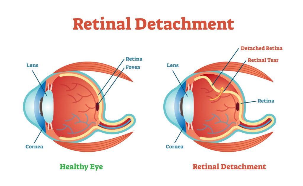
Retinal detachment occurs when the retina moves from its normal position at the back of the eye. The retina is a thin layer of tissue that processes light so you can see.

A detached retina is a medical emergency that requires immediate care. Call your eye doctor right away if you experience any of these warning signs of retinal detachment:
- A sudden increase in eye floaters
- Seeing flashing lights in one or both eyes (photopsia)
- A dark shadow or curtain on the sides or center of your visual field
Retinal cells cannot get the nourishment and oxygen needed from blood vessels when detachment occurs. Early treatment can help prevent irreversible loss of vision.
Symptoms of Retinal Detachment
Retinal detachment is painless. Don’t assume that lack of discomfort or pain means there isn’t a problem.
Symptoms of retinal detachment include:
- The sudden appearance of dark specks in your field of vision (floaters)
- Flashes of light in one eye or both
- Reduction in peripheral vision
- Blurred vision
- A dark, curtain-like shadow that slides over your field of vision
Seek immediate medical attention if you experience any of the above symptoms.
What Causes Detached Retinas?
The cause of retinal detachment depends on the type you have:
Rhegmatogenous
These are the most common type. Rhegmatogenous detachments result from a hole or tear in the retina.
A retinal tear allows fluid to pass into and collect beneath the retina. Gradually, the retina pulls away from the back of the eye, causing blood loss and decreased vision.
What Causes Rhegmatogenous Detached Retinas?
Age is the most common cause of rhegmatogenous retinal detachment. As someone ages, the vitreous gel in their eyes shrinks or becomes more liquid.
Posterior vitreous detachment (PVD) is when the vitreous separates from the retina. This normally occurs without complications. A retinal tear is one complication that can lead to detachment.
Other risk factors for rhegmatogenous retinal detachment include:
- Eye injury
- Eye surgery
- Myopia (nearsightedness)
Tractional
A tractional detachment is caused by the growth of scar tissue on the retina’s surface that pulls at the retina.
What Causes Tractional Detached Retinas?
Diabetic retinopathy is the most common cause of tractional detachment. This eye condition affects people with poorly controlled diabetes.
Diabetic retinopathy damages the retina’s blood vessels, causing scar tissue to form on the retina.
Other causes of tractional detachment include:
- Eye diseases
- Eye infections
- Eye swelling
Exudative
This type of detachment happens when fluid accumulates behind the retina, but there isn’t a hole or tear. As more fluid accumulates, it can cause separation and detachment.
What Causes Exudative Detached Retinas?
Leaking blood vessels or swelling in the back of the eye are the most common causes of exudative retinal detachment.
Swelling and leaking blood vessels can be caused by:
- Age-related macular degeneration (AMD)
- Eye trauma or injury
- Tumors in the eye
- Inflammatory disorders
Who is at Risk for Retinal Detachment?
Anyone can experience retinal detachment, but some people have a higher risk.
Risk factors for retinal detachment include:
- Advanced age (50 and older)
- Family history of retinal detachment
- Extreme myopia (nearsightedness)
- Previous retinal detachment in one eye
- Previous eye surgery, especially cataract surgery
- Eye diseases, including uveitis, lattice degeneration, retinoschisis
- Previous severe eye injuries
- Diabetes
How Is Retinal Detachment Diagnosed?
A doctor must diagnose and treat retinal detachment. If the examination reveals no holes or tearing despite your symptoms, your doctor will schedule a follow-up visit.
If you experience any additional or new symptoms before the follow-up visit, it’s important to contact your doctor.
Tests used to diagnose detachment include:
Dilated Eye Exam
If your eye doctor suspects you have a detached retina, they’ll perform a dilated exam. They’ll give you special drops to widen your pupils, then examine the back of your eye with a bright light.
Ultrasound
If there is bleeding in the eye that makes it difficult to examine the retina, your doctor might order an ultrasound to get a better view of the back of the eye.
Treatment Options for Detached Retinas
If the examination reveals detachment, your doctor will order surgery within hours or days of the exam.
There are three surgical procedures used to repair detachment, including:
Pneumatic Retinopexy
This procedure involves injecting a gas bubble into the vitreous cavity in the center of the eye. The bubble pushes the damaged part of the retina against the eye’s wall, which stops the flow of fluid behind the retina.
Once they’ve halted the flow, the surgeon uses cryopexy or laser to repair the retinal tear. The bubble of air or gas and any liquid is absorbed into the eye, allowing the retina to adhere back into place.
Scleral Buckling
Scleral buckling uses a silicone material that is sewn in place by the surgeon to cover the sclera (white of the eye). It creates an indentation in the wall of the eye and relieves the tugging and pulling on the retina by the accumulated vitreous liquid.
The silicone cover doesn’t block vision and can remain in place forever. If there are multiple holes, the surgeon can attach covers that encircle the eye with support.
Vitrectomy
This procedure drains the accumulated vitreous and uses air, gas, or silicone oil to fill the vitreous space and flatten the retina. The injected material is eventually absorbed, and the space refills with fluid.

Sometimes, the fluid must be surgically removed several months after the procedure. Vitrectomy is often used in combination with scleral buckling.
Can a Retinal Detachment Heal on Its Own?
No. Left untreated, retinal detachment worsens and can lead to permanent vision loss.
Retinal detachment must be treated by a medical professional and requires surgery to correct. This is the only way to reattach the retina so it can receive the blood supply it needs to remain healthy.
How to Prevent Retinal Detachment
There is no way to prevent retinal detachment. However, there are some things you can do to reduce your risk, including:
- Get regular eye exams. This is especially important if you have nearsightedness. Myopia increases your risk for retinal detachment.
- Protect your eyes. Use protective eyewear when playing sports or engaging in activities that might lead to an eye injury.
- Get prompt treatment. Seek medical attention as soon as possible if you notice symptoms of a detached retina.
Outlook
The outlook for retinal detachment depends on several factors, including:
- How good your vision was before the retina detached
- The severity of the detachment
- Whether any complications arose
Your eye doctor will discuss the type of vision improvement you can expect. Retinal detachment surgery has a high success rate. The repair works in about 90% to 95% of cases.3
Summary
Retinal detachment is a medical emergency. It occurs when the light-sensitive membrane separates from the back of the eye.
Warning signs of a detached retina include increased floaters and light flashes. Prompt treatment is necessary to prevent permanent vision loss.
Common causes of a detached retina include retinal tears, eye injuries, and diseases like diabetic neuropathy and macular degeneration.
In this article


